

Scientists from Scripps Research report that they have developed a novel method to examine how proteins interact with drug-like small molecules in human cells—revealing critical information about how to potentially target them therapeutically.
Their study, “Enhanced mapping of small-molecule binding sites in cells,” published in Nature Chemical Biology, uses a combination of chemistry and analytical techniques to reveal the specific places where proteins and small molecules bind together. Ultimately, this method could lead to the development of more targeted and effective therapeutics.
“Our new technology could be used to find new druggable sites on proteins for any human disease, from cancer to Alzheimer’s disease,” said Christopher Parker, PhD, associate professor, department of chemistry, and senior author of the study. “We’re unrestricted in how this could be used. Our work has the potential to usher in a whole new way of drug discovery.”
The Parker lab aims to discover how proteins function in every human cell type to develop effective therapeutics for a wide range of human diseases. In this study, Parker and his team built off his initial work in the lab of Scripps Research professor Benjamin Cravatt, PhD, to create a novel method of examining how proteins interact with small molecules in living cells.
Analytical strategy
The team developed an analytical strategy to better understand how these proteins engage with small molecules at much higher resolution than ever before. To do this, they used chemical photoaffinity probes, which are molecules that can be activated by light to allow the probes to capture a bound protein.
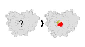
By gathering data from the interactions of proteins with photoaffinity probes, the Parker team identified locations on proteins where small molecules could connect and bind. Essentially, the team found over a thousand new locks (binding sites on the proteins) and corresponding keys (small molecules), the vast majority of which were new places of small-molecule binding that had not been reported before.
Additionally, they found new features of the binding sites—such as new shapes.
“Identifying these specific binding sites will help scientists design new molecules that fit these pockets even better, potentially leading to more effective therapeutics,” said Jacob M. Wozniak, PhD, co-first author, and former postdoctoral fellow in the Parker lab. The other co-first author of the paper was Weichao Li, PhD, a research associate also in the Parker lab.
Using the data in this study and collaborating with co-author Stefano Forli, PhD, associate professor in the department of integrative structural and computational biology, the authors then modeled how certain molecules might bind to these proteins. This library of information could be used to design therapeutics that interact with proteins in a more targeted way, according to the researchers.
“Our new process reveals additional opportunities for therapeutic intervention and discovery in human cells,” said Parker. “Next, we plan to use this technology to target proteins relevant for autoimmune diseases and cancer.”
The post Illuminating Druggable Sites on Proteins appeared first on GEN – Genetic Engineering and Biotechnology News.


